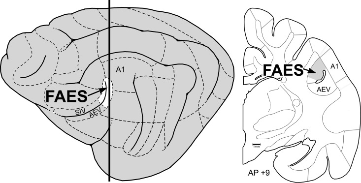
Figure 1.
Location of the auditory FAES. The lateral view of the cat cortex (left) shows the location of the anterior ectosylvian sulcus, depicted as opened to expose its dorsal and ventral banks. The posterior banks (white, at arrow) contain the auditory representation of the FAES, whereas the anterior-dorsal bank contains the SIV and the anterior-ventral bank contains the AEV. Dashed lines indicate functional subdivisions of the cortex, where primary auditory cortex (A1) is labeled as a point of reference. The thick vertical line indicates the approximate anterior–posterior (AP) level from which the coronal section (right; ∼AP + 9) was taken. Here, the anterior ectosylvian sulcus resides deep to the middle ectosylvian gyrus (labeled A1) and contains the FAES (at arrow). Gray lines depict the functional subdivisions of the cortex; the grayed-area of the coronal section represents the smaller representation of the FAES region in early deaf cats (after Wong et al. 2014).

