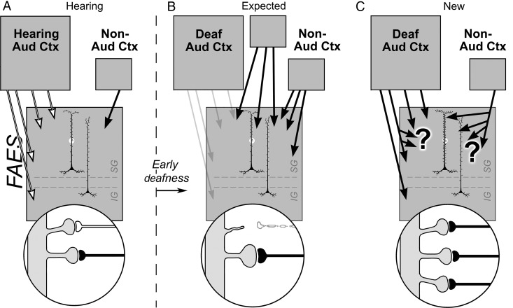
Figure 10.
Summary of synaptic basis for deafness-induced cross-modal plasticity in the FAES. For hearing animals (A), neurons in the infraganular (IG) and supragranular (SG) laminae of the FAES (large gray box) predominantly receive inputs from auditory cortex (multiple white arrows) as well as a small projection from non-auditory cortical regions (black arrow). The highlighted segment of an FAES neuron's dendritic shaft is enlarged (bottom circle) to show spines with synaptic inputs from auditory cortex (white axon) as well as from non-auditory cortex (black axon). The schematics to the right of the vertical dashed line represent changes in the FAES induced by early deafness. (B) The historically expected effects of cross-modal plasticity represented by increases in inputs from new or existing non-auditory areas (black arrows) and/or by increased synaptic weighting/efficacy of existing non-auditory inputs (enlarged black axon and terminal). Although not explicitly stated, it has also been assumed that inputs from auditory cortex are lost/reduced after early deafness (retracted spine and white axon; faded arrows). In (C), recent data have observed that increased projections from new or existing non-auditory areas were not found, nor were connections with auditory cortex lost. In addition, dendritic spine numbers increased, without increases in spine size. These observations imply that increases in spine density may be paralleled by increases in afferent branching (large “?”) and that most inputs to the FAES (from either auditory or non-auditory regions) now relay non-auditory signals (black axons).

