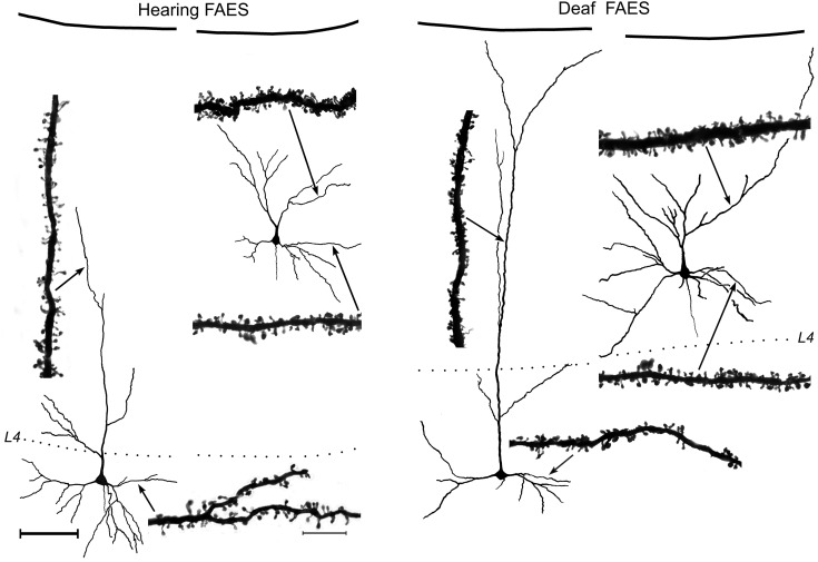
Figure 2.
Golgi-stained pyramidal neurons from hearing (left) and early deaf (right) FAES are reconstructed relative to supragranular or infragranular laminar location (pia = top; dotted line = granular layer L4) using camera lucida. Photomicrographs (×1000, oil) depict the dendritic spines of selected examples of apical and basilar dendrites at sites indicated by the arrows. Scale bars for the neurons and dendrites are shown in the left panel: neuron scale bar (left) = 100 μm; dendritic segment scale bar = 10 μm.

