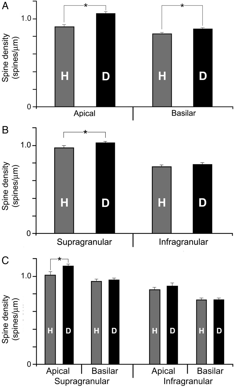
Figure 5.
Only specific dendritic branches of FAES neurons from early deaf cats exhibit higher dendritic spine density than their hearing counterparts. (A) The bar graph shows that the average (±SE) dendritic spine density of apical or basilar dendritic segments was significantly higher (“asterisk,” t-test, P < 0.012) in early deaf animals than in the hearing controls. In contrast, in (B), the bar graph (mean ± SE) shows that the average dendritic spine density of FAES neurons located in the supragranular layers was significantly higher (t-test, P < 0.012) in early deaf animals than in the hearing controls, but not in infragranular neurons. Furthermore, the bar graph (mean ± SE) in (C) divides the data into apical/basilar segments based on neuronal laminar location. When sorted by lamina, only apical dendrites of supragranular neurons showed a significant increase in spine density in early deaf animals. The other categories (supragranular–basilar segments; infragranular apical segments, and infragranular basilar segments) did not reveal significant alterations in spine density within the different treatment groups.

