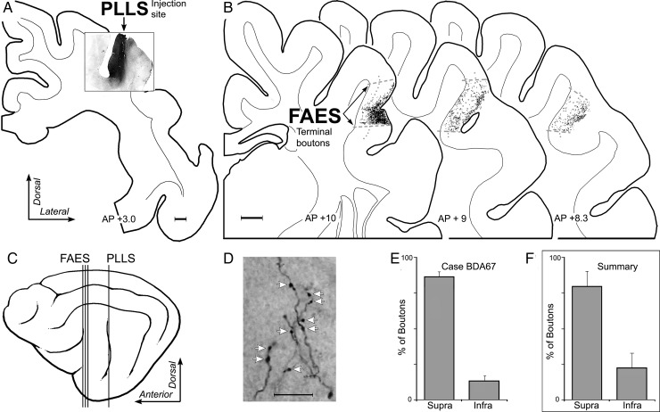
Figure 7.
Laminar termination in the FAES of non-auditory inputs from visual PLLS. In (A), the coronal section shows the injection site (black area) in the lateral bank of the suprasylvian sulcus corresponding to the location of visual area PLLS (scale = 1 mm). In (B), coronal sections through the anterior (left), middle, and posterior (right) regions of the FAES (borders indicated by dashed gray lines; layer 4 indicated by dotted gray line) displaying terminal boutons (1 black dot = 1 bouton; scale = 1 mm) labeled from the PLLS injection site. The lateral view of cortex (C) illustrates the location (vertical lines) of the coronal sections through the PLLS (shown in A) and FAES (B). (D) is a micrograph (×1000, oil; scale = 10 μm) taken of representative PLLS-labeled axons and boutons (indicated at white arrows) within the FAES. When the number of boutons labeled from the PLLS were counted within the supragranular versus infragranular layers of the FAES for this case, the overwhelming proportion (mean = 86% ± 3 SE) was identified within supragranular layers, as quantitatively summarized in the bar graphs (E). Similarly, (F) the levels of boutons labeled from PLLS for all cases and sections predominantly (mean = 78% ± 10 SE) terminated within the supragranular layers of the FAES.

