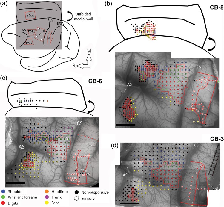
Figure 1.
Electrophysiological mapping. (a) Cartoon showing a lateral view of the brain and the approximate location of the craniotomy (gray square) and the areas of the frontal and parietal cortex studied with electrophysiological mapping methods. The medial wall is shown unfolded (curved arrow). (b) Motor and sensory mapping data in CB-8. Each colored dot on the digital photograph of the cortex represents a microelectrode penetration site. The evoked movement (frontal cortex) or somatosensory receptive field location (parietal cortex) is color-coded (legend). In the parietal cortex, the same color-code is used for the receptive field location on the body, but the dots have white contours. The hand representations of M1, PMv, PMd, and SMA are outlined with red contours. In the parietal cortex, the medio-lateral extent of the hand representation in S1 and the location of rostro-caudal borders between area 1, area 2, and area 5 are shown with red lines. Note that the location of stimulation sites for SMA are approximated based on the distance from PMd and the depth of the electrode. They are shown over a cartoon representation of the unfolded medial wall to provide an estimated location of the sites in relation to the medial wall's convexity and the cingulate sulcus. The location of these 2 landmarks is based on the anatomical reconstructions of each monkey. (c) Motor and sensory mapping data for CB-6. In this animal, we were able to locate SMA but got limited data from it. As for CB-8, the location of the stimulation sites in SMA is estimated. In S1, we did not physiologically define the medial border of the hand representation. Based on other animals, we estimated the location of this border and show it with a dotted red line. (d) Motor and sensory mapping data for CB-3. In this animal, we did not locate SMA. AS: arcuate sulcus; CS: central sulcus; IPS: intraparietal sulcus; M1: primary motor cortex; PMd: dorsal premotor cortex; PMv: ventral premotor cortex; SMA: supplementary motor area; 1: area 1; 2: area 2; 5: area 5; M: medial; R: rostral. Scale bar = 5 mm.

