Figure 3.
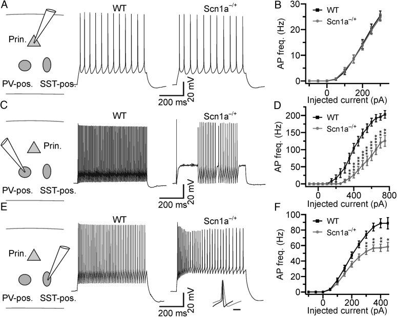
Cell type-specific dysfunction of excitability in brain slices from Scn1a−/+ mice. (A) Left: schematic of the experimental configuration. Prin., principal neuron; PV-pos., parvalbumin-positive interneuron; SST-pos., somatostatin-positive interneuron. Right: representative current-clamp recordings from pyramidal neurons recorded from WT (left) and Scn1a−/+ (right) mice in cortical slice. (B) Average frequency of AP firing in principal neurons as a function of injected current for WT (N = 27 cells, black) and Scn1a−/+ (N = 27 cells, gray). (C) Representative current-clamp recordings from Tomato-positive interneurons recorded in cortical slices from a WT-PV-Tomato (WT, left) and a Scn1a−/+-PV-Tomato (Scn1a−/+, right) mouse. (D) Average frequency of AP firing as a function of injected current for fluorescent cells recorded in WT-PV-Tomato mice (N = 21, black) and Scn1a−/+-PV-Tomato mice (N = 28, gray). The asterisks indicate significance at each value of current injection evaluated with the Bonferroni post hoc test. In this as well as in other figures: *P < 0.05; **P < 0.01; ***P < 0.001. (E) Representative current-clamp recordings from Tomato-positive cells recorded in cortical slices from WT-SST-Tomato (left) and Scn1a−/+ -SST-Tomato (right) animals. The inset shows the overlap of one AP recorded in a PV-positive cell from a Scn1a−/+-PV-Tomato mouse and one AP recorded in a SST-positive cell from a Scn1a−/+-SST-Tomato mouse. Traces are normalized to the AP maximal amplitude. Scale bar 10 ms. (F) Average frequency of AP firing as a function of injected current for cells recorded in WT-SST-Tomato animals (N = 25, black) and Scn1a−/+ -SST-Tomato animals (N = 28, gray).

