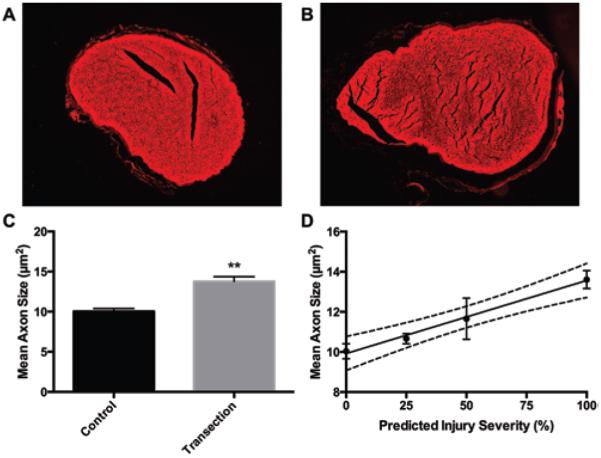
FIG. 1.
Predicted nerve injury severity correlated with proximal axon caliber. A and B: Representative fluorescent microscopy images of CM-DiI membrane label in (A) sham and (B) completely transected sciatic nerves. Axoplasm is visible as holes in the center of red membrane rings. C: Axon caliber was measured 3 mm proximal to the injury site for all axons visible in the axial cross-sections of sham and completely transected nerves (mean ± SEM; n = 6). D: Mean axon caliber correlated moderately with the predicted injury severity (r2 = 0.59; n = 15; p = 0.002). However, correlation strongly improved after removing the nerves predicted to be 75% transected (r2 = 0.82; n = 12; p = 0.0001). Dashed lines indicate the 95% CI. **p < 0.01, unpaired t-test with Welch correction.

