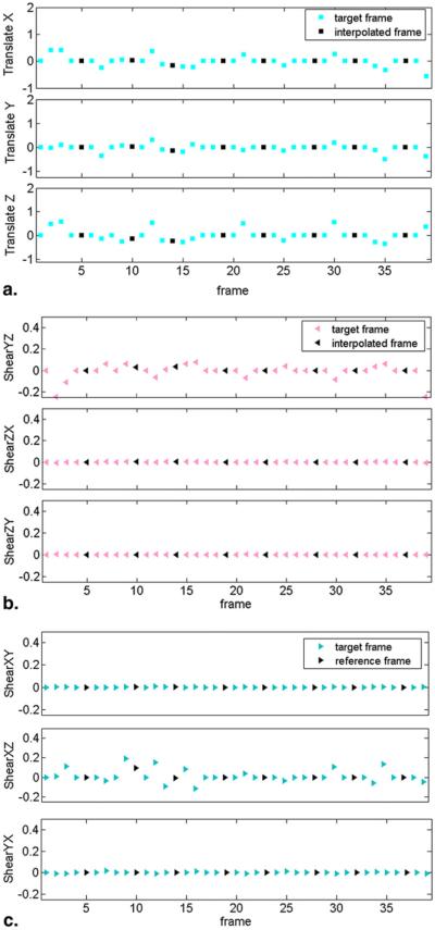
Figure 3.
Plot of three-dimensional affine transformation parameters for the 39 frames acquired during an abdominal exam. (a) Translational parameters (translate X, translate Y, and translate Z), (b) Shear parameters (shear XY, shear XZ, and shear YX) and (c) (shear YZ, shear ZX, and shear ZY). Motion parameters were estimated during acquisition of high-signal low b-value images (colored markers represent target frames), which can then be interpolated to the adjacent high b-value images that lack sufficient signal for reliable motion estimation (black markers represent interpolated frames).

