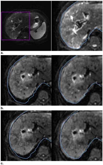
Figure 5.
Registration of target image to the reference image. (a) Reference image (frame 1, b = 0 s/mm2). For comparison, the contour of the liver (blue) from frame 1 is copied in its original location in each subsequent frame. (b) Pre- (left) and postregistration images of the same slice from frame 29 (b = 70 s/mm2). In the preregistered image, the liver and the contour do not match. Postregistration, there is an improvement in the alignment of the liver edge and the contour copied from frame 1. (c) Similarly, for frame 36 (b = 150 s/mm2), the liver edge in the preregistered image (left) does not match the contour. The alignment is improved in the postregistration image (right).

