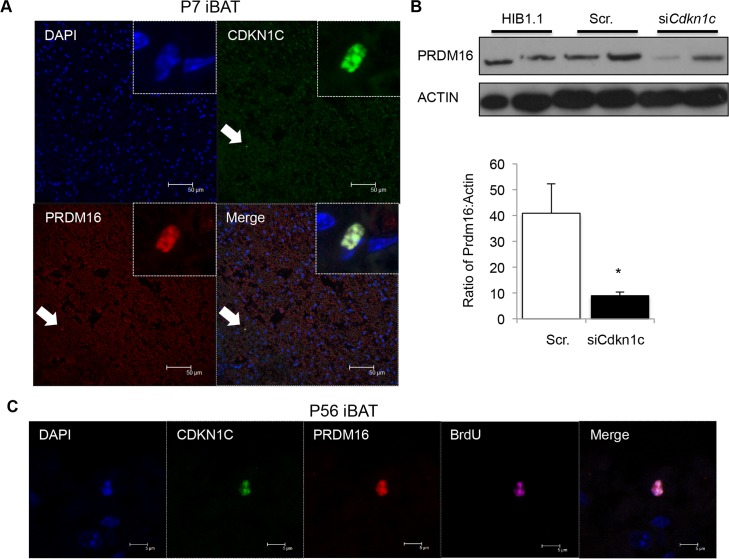
Fig 8. CDKN1C and PRDM16 co-localise to the nucleus of rare BrdU label-retaining cells in iBAT.
(A) Confocal imaging of P7 iBAT co-stained for CDKN1C and PRDM16. DNA is stained with 4′,6-diamidino-2-phenylindole (DAPI, blue). (B) Western analysis of PRDM16 protein after siRNA-induced knock-down of Cdkn1c in the undifferentiated brown fat preadipocyte cell line HIB1.1. (C) Immunohistochemistry for CDKN1C (green), PRDM16 (red) and BRDU (purple) in WT iBAT 8 weeks after in utero pulsed exposure to BrdU. DNA (DAPI, blue).

