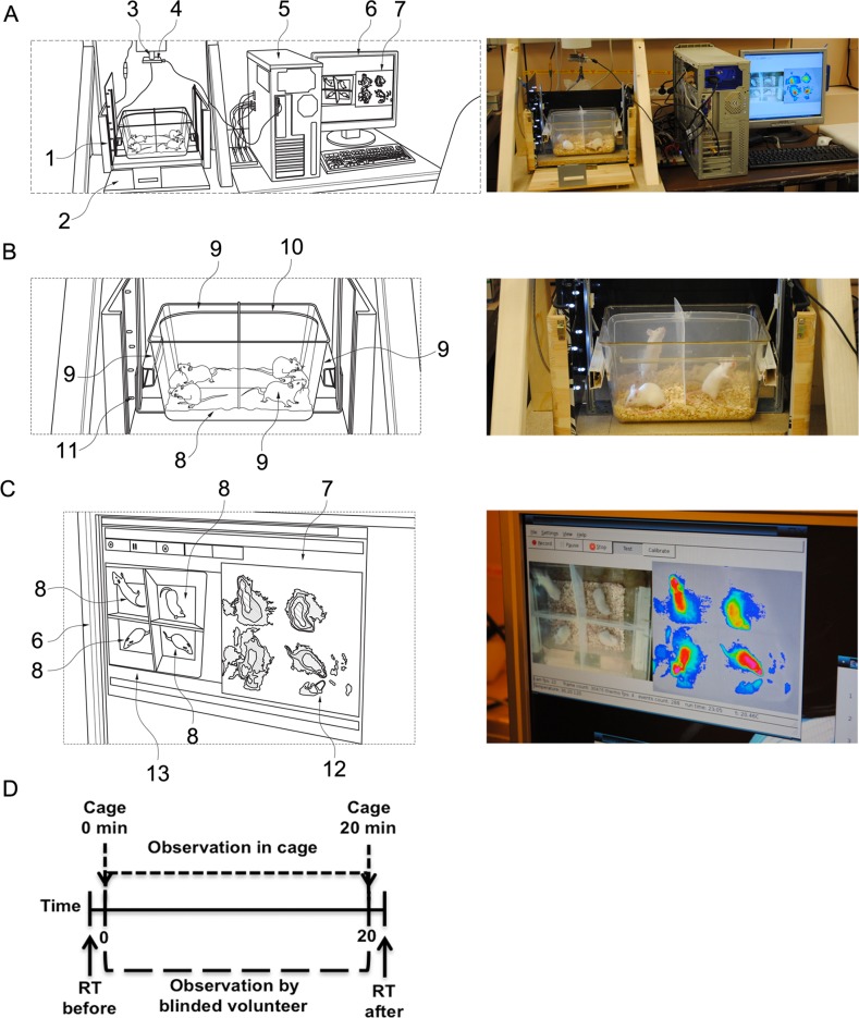
Fig 1. The schematic (left panels) and real set up (right panels) of the imaging cage.
(A) The complete device is composed of a type II animal cage, a photo- and a thermo-camera, which are connected to a computer and software. (1) Outer box. (2) Outer sidewall. (3) Above the cage and height-adjustable, a heat camera or heat image camera (false colour infrared camera or infrared camera) is fixed with a reference heat electrode placed in the field of view for precise calibration against a reference temperature module. (4) Mounted true colour camera, which records live real image pictures during the experiment. (5) Data are recorded and processed, and translated by software to a personal computer for processing, identifying grey scales in video input from connected infrared camera 3. The software comprises a data processing part and (7) a graphical user interface, which is commonly displayed on (6) a screen. (B) Four small animals, in this case mice, can be placed and monitored at the same time in the cage. (8) Transparent enclosure adapted to house four mice. (9) The cage comprises four outer sidewalls and (10) two inner walls, as well as a bottom portion. (11) Vertical movements can be additionally recorded to the imaging data by photo sensors comprising transmitters and receivers. (C) The mice are individually monitored, where the thermo measurements and the live video from the cage appear at the same time on the monitor. (12) False colour screen output. Two pictures are recorded and may be visualized on screens. The software translates the grey image recordings from the infrared camera (Fig 1A-3) in real-time into a false colour screen output. (13) True colour image of the true colour camera is displayed in parallel. (D) Rectal temperature (RT) was measured manually before and 20 minutes after i.v. allergen challenge of hypersensitive or negative control mice. Immediately after the challenge, the same mice were placed into the imaging cage (0 min), and the cameras monitored surface body temperature and movement activity continuously for 20 minutes (20 min).

