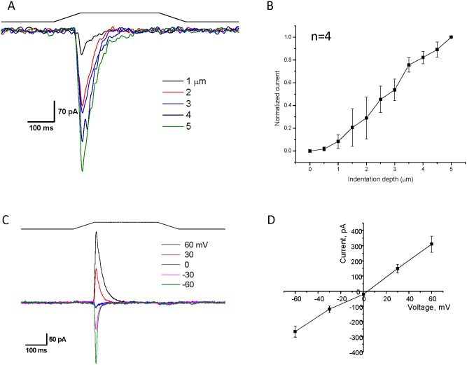
Fig 2. Two hPIEZO1 segments reassemble into a functional channel.
A plasmid allowing for the expression of two protein segments (1–1591 with a mCherry protein attached to its C-terminal and 1592–2521 with an N-terminal GFP) was transfected into HEK293T cells. Cells were identified by both green and red fluorescent indicating that both parts of the protein were expressed. Panel A shows whole cell currents elicited with a fire-polished probe that mechanically stimulates the cell by pressing on the membrane (holding potential = -60 mV). The black line indicates the stimulus pulse. Panel B demonstrates that increasing depth of penetration produces more current (n = 6, error bars are S.D.) Panel C is a cell-attached current trace at the indicated voltages, and Panel D is a plot of the current versus voltage showing that the channel reverses around 0mV and is similar to the wild type channel (n = 4. error bars are S.D).

