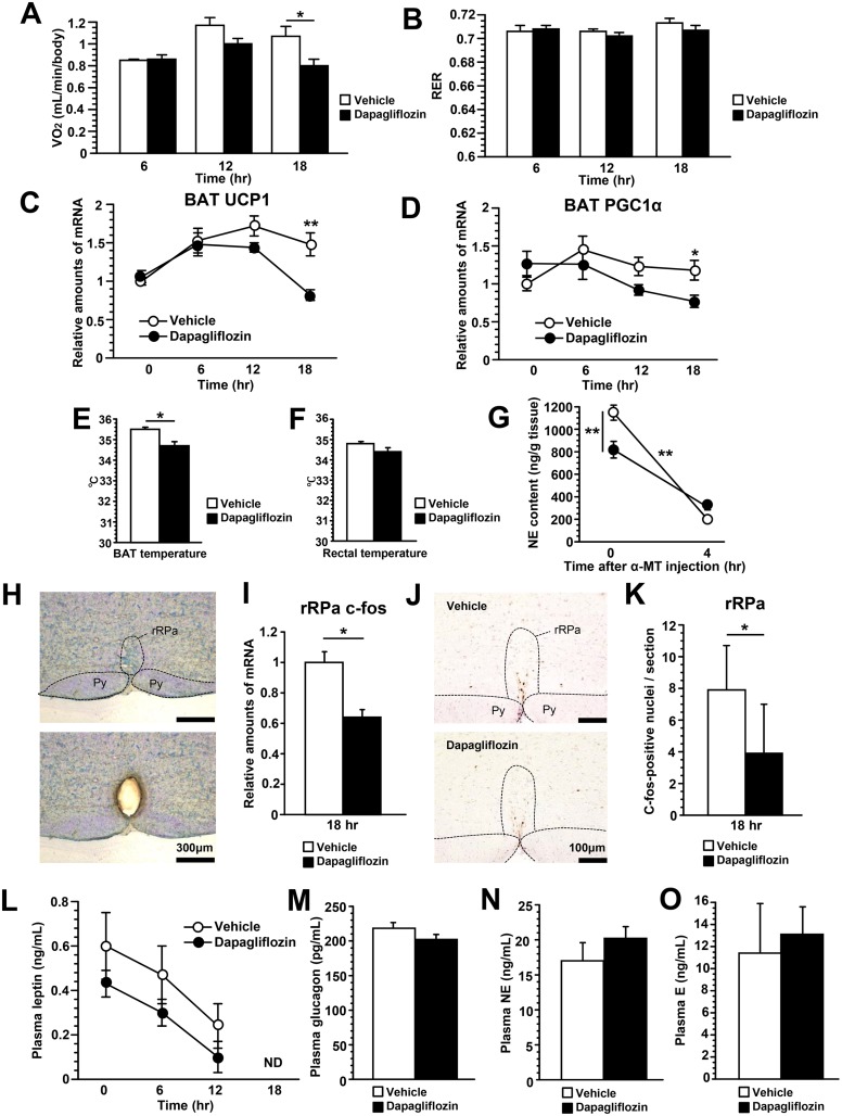
Fig 2. Dapagliflozin acutely suppressed BAT thermogenesis by reducing sympathetic nerve activity.
Oxygen consumption during the 18 hours following dapagliflozin administration (A). Respiratory exchange rate (RER) during the 18 hours following dapagliflozin administration (B). Relative amounts of UCP1 (C) and PGC1α (D) mRNA in BAT. BAT (E) and rectal (F) temperatures 18 hours after dapagliflozin administration. NE turnover in BAT 18 hours after dapagliflozin administration (G). The difference in the NE decline between SGLT2i- and control-mice due to α-methyl-p-tyrosine (α-MT) treatment was statistically analyzed employing two-way ANOVA. Coronal section of the rostral ventromedial medulla before (upper panel) and after (lower panel) microdissection of the rRPa. Py, pyramidal tract (H). Relative amounts of c-fos mRNA in the rRPa 18 hours after dapagliflozin administration (I). Immunohistochemical detection of c-fos protein in the rRPa (J). The number of c-fos positive neurons in the rRPa (K). Plasma leptin levels during the 18 hours following dapagliflozin administration (L). Plasma glucagon (M), NE (N) and epinephrine (E) (O) levels 18 hours after dapagliflozin administration. Values shown are means ±SEM (n = 6–7 per group). Significance as compared to control-mice is indicated (**P < 0.01 and *P < 0.05 and). ND = not detectable.

