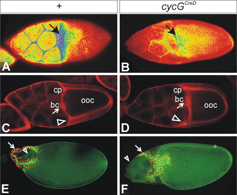
Fig 7. Notch expression and nurse cell positioning are affected in cycG mutant ovaries.
(A, B) Superficial view onto a stage 10 egg chamber stained for Notch protein. Shown is a false colour picture with colours representing strength of signal from red (low) to yellow (intermediate) to blue (strong). Pictures were taken with identical settings from ovaries stained in parallel experiments with identical conditions. (A) In the wild type, Notch is expressed in a graded fashion and is mostly enriched in the prospective floor cells of the developing dorsal appendages (arrow). (B) In the CycGCreD homozygous mutant, Notch expression is markedly reduced in the entire follicle (arrow). (C, D) A sagittal view shows the enrichment of Notch protein in the centripetal cells (cp; arrowhead) and the border cells (bc; arrow) at the anterior border of the oocyte (ooc). The anterior border is slightly convex or straight in the wild type (C). (D) In the cycGCreD homozygote, the nurse cells push into the oocyte, i.e. the centripetal cells (cp; arrowhead) are located anterior relative to the border cells (bc; arrow). (E, F) A superficial view onto stage 13–14 egg chambers is shown. The developing dorsal appendages (arrow) are marked with anti-Broad antibodies (nuclear, red); the green staining of the eggshell is due to auto-fluorescence. In the wild type (E), the nurse cells have been eliminated by apoptosis (arrowhead). (F) Egg chambers of homozygous cycGCreD females frequently show a ‘dumpless’ phenotype, because extant nurse cells are found outside of the oocyte beyond stage 14 (arrowhead).

