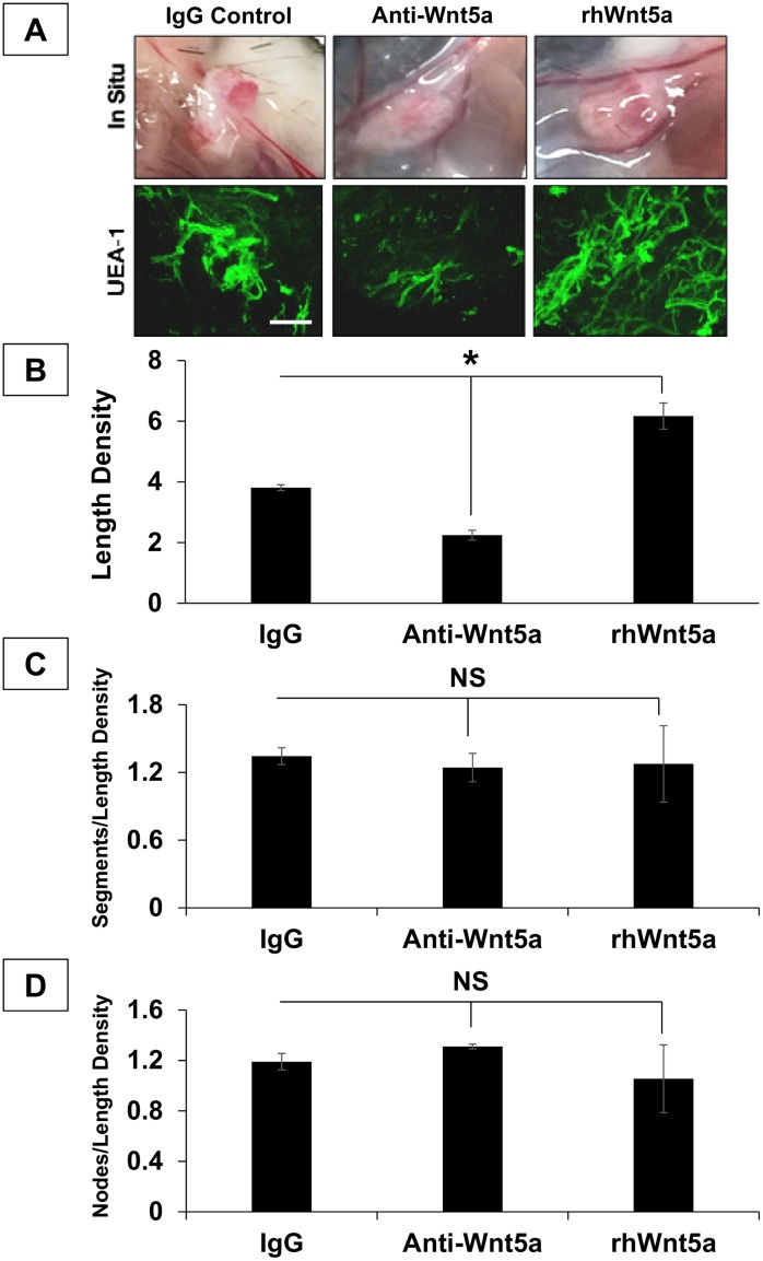
Fig 6. Wnt5a drives hSVF EC microvascular assembly in vivo.
(A) 3D collagen-I constructs containing hSVF an IgG isotype control, Wnt5a neutralizing antibody (anti-Wnt5a), or rhWnt5a (3 animals per treatment; 1 representative donor cell line was used) were implanted for 2w. Images show 2w constructs in situ. Constructs were then labeled with UEA1 (green) and imaged by confocal microscopy. Scale bar = 20x, 100μm. (B) Anti-Wnt5a treatment significantly reduced the length density compared to the IgG control (*p ≤ 0.05), while rhWnt5a addition significantly increased this ratio (*p ≤ 0.05). (C) Normalization of segment number to length density showed no significant affect with either the anti-Wnt5a or the rhWnt5a treatments compared to IgG controls. (D) The ratio of node number to length density also showed no significant difference between the IgG control, the anti-Wnt5a treatment, and treatment with rhWnt5a. NS = not significant. Bars shown as mean ± S.E.M.

