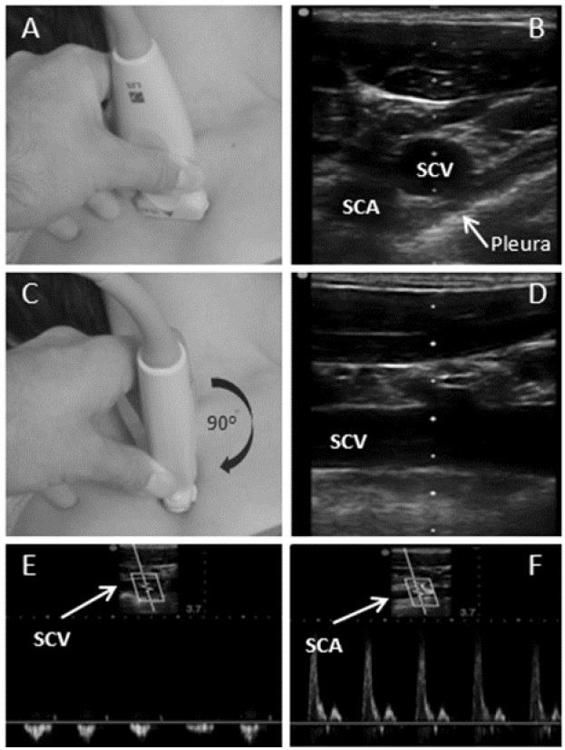
Figure 2.

A) Linear transducer is placed perpendicularly and inferior to clavicle. B) Identified anatomical structures include the transverse (short axis) view of subclavian vein (SCV), subclavian artery (SCA) and pleura. C) With SCV centrally positioned, the transducer is rotated 90° clockwise until D) longitudinal view of subclavian vein is obtained. E) Pulse-wave Doppler view of the SCV confirms non-pulsatile flow and identifies the vessel. F) Tilting the transducer cephalad enables the visualization and identification of SCA with pulse-wave Doppler ultrasound for better anatomic orientation.
