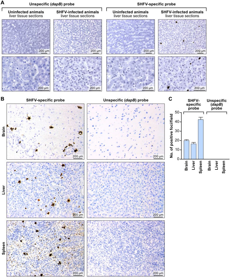
Fig 3. In situ detection of SHFV RNA from tissue sections from an SHFV-infected rhesus monkey using RNAscope® in situ hybridization.
(A) Liver sections from an uninfected or SHFV-infected rhesus monkey labeled with unspecific (dapB) or SHFV-specific target probes. Top: all images were originally taken at 200X magnification. Bottom: all images were originally taken at 400X magnification. Positive results manifest as brown staining. (B) Detection of SHFV RNA in brain, spleen, and additional liver sections of the same animal (original magnification 400X). Positive results manifest as brown staining after amplification. (C) Quantification of SHFV RNA-positive foci in brain, liver, and spleen sections by counting; four fields were counted per tissue section of 200X-magnified images (p value calculated by multiple t-test analysis with GraphPad Prism 6 software).

