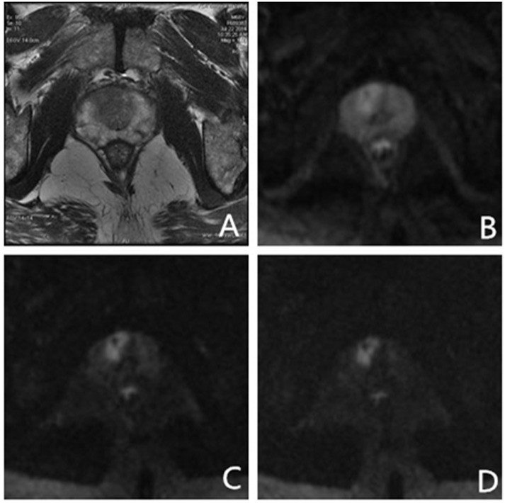
Fig 5. Images of a 68 yr old male patient with total PSA 10.5 ng/ml and free PSA 1.66 ng/ml. Systematic transrectal biopsy confirmed PCa (Gleason score 4 + 3 = 7).
(A) High-solution T2-weighted imaging shows low signal intensity area in the right TZ, and PI-RADS score of 2 due to subtle mass effect; (B) PI-RADS score of 3 on DWI with b-value 1000 s/mm2. The cancer is ambiguous because of high signal intensity from surrounding parenchyma; (C, D) PI-RADS score of 5 on ultrahigh DWI with b-value 2000, 3000 s/mm2. High signal intensity areas in the right portion of the TZ are clearly visible. Key: PSA—Prostate specific antigen; PCa—prostate cancer; TZ—transition zone; DWI—diffusion-weighted imaging; PI-RADS—Prostate Imaging Reporting and Data System.

