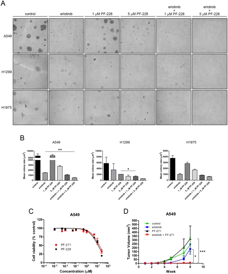
Fig 6. Combination treatment with erlotinib and FAK inhibitor is more effective in reducing colony growth in vitro and tumor growth in vivo.
(A) 3-dimensional colony formation was assessed from NSCLC cells embedded in growth factor reduced BME in 4-well chamber slides in the presence of either vehicle control (DMSO), erlotinib (10 μM), PF-228 (1 μM or 5 μM), or the combinations of erlotinib and PF-228 in complete media. Media with fresh drug was replenished every 3 days and colony size was assessed after 13 days. Images representative of two independently performed experiments are shown. (B) Mean colony area ± SEM is presented with statistically significant differences in mean colony size determined by unpaired Student’s t tests (** P = 0.0025, A549; * P = 0.044, H1299) from two independently performed experiments. (C) FAK inhibitors PF-228 and PF-271 show similar effectiveness in reducing cell viability in vitro. A549 cells were treated with either inhibitor for 48 hours and cell viability was assessed by MTT assay. Data indicates the mean ± SEM for log[inhibitor] vs. normalized response from two independently performed experiments. (D) In vivo data was collected from CD-1 nude mice injected subcutaneously with A549 cells and treated by oral gavage beginning 3 days post tumor cell injection. Data presented is the mean ± SEM for tumor volume from 4 mice per treatment each with cells injected into both hind flanks (n = 8 tumors/treatment), with statistically significant differences in tumor volume at week 8 determined by ANOVA (* P < 0.05, *** P < 0.001).

