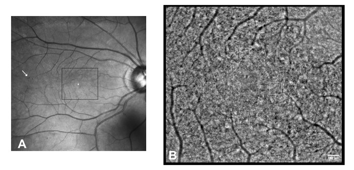
Fig 1. Adaptive optics montage of the central retina in the right eye of a patient with a diagnosis of type 1 diabetes mellitus in the past 37 years (DM1_P7).
A) Wide field fundus image. The arrow highlights a cluster of spot hemorragies outside the region of interest. B) Adaptive optics montage of the central retina showing the photoreceptor mosaic (2.46x2.18 mm). Analysis of the cone mosaic was done in 160x160 μm sampling areas on four locations at 1.5 degrees eccentric from the fovea along all retinal meridians. Scale bar: 160 μm.

