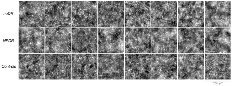
Fig 4. Adaptive optics images of the photoreceptor mosaic for all patients with diabetes and eight representative age-matched controls acquired at 1.50 degrees superior from the fovea.
The upper row includes the eyes with noDR (from DM1_P9 to DM1_P16 from the left to right respectively), the middle row the eyes with NPDR (from DM1_P1 to DM1_P8 from the left to right respectively) and the lower row the control eyes (C30, C32, C33, C37, C39, C41, C44 and C48 from the left to right respectively). Each window is 160x160 μm. The cones were reliably identified in all AO images collected from study cases and controls.

