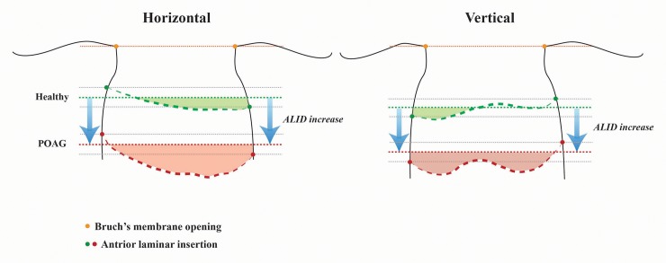
Fig 5. The schematic diagram showing the structural difference of lamina cribrosa (LC) between the eyes with primary open-angle glaucoma (POAG) and healthy individuals.
Structural differences of the LC between POAG and healthy eyes are demonstrated. The orange dotted line corresponds to the reference plane of Bruch’s membrane openings. The thicker green and red dotted line indicates the anterior LC surface of healthy and POAG eyes, respectively. The thinner green and red dotted line presents the level of mean anterior laminar insertion depth (ALID). The green and red shaded area represents the relative posterior location of the anterior LC according to the mean ALID level of healthy and POAG eyes, respectively. Each shaded area reflects the degree of LC posterior bowing (LC curvature index). The ALID and LC posterior bowing (LC curvature index) are higher in POAG eyes than in healthy eyes. Note that vertical insertions are deeper than horizontal insertions. Additionally, the horizontal LC curvature index is larger than the vertical LC curvature index in both healthy and POAG eyes, due to the central hump-like structure in the vertical scan.

