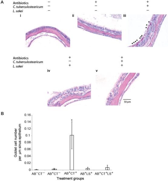
Fig. 4.
(A) L. sakei protects sinus mucosa from C. tuberculostearicum–induced pathogenesis. PAS-stained histological sections of murine sinuses representative of animals in each treatment group at ×60 magnification (panels i to v). Triplicate views of maxillary sinuses from two mice per treatment group were used to determine physiology (representative images are shown). (B) Enumeration of goblet cells across murine treatment groups (n = 3 animals per group). Values represent means ± SEM.

