
Figure 4. p63 protein expression in SOD1(G86R) muscle.
(A) Proteins from muscles were immuno-precipitated with a p63 antibody and then separated on a 10% SDS PAGE gel. Western blot experiment was performed using an antibody against TAp63. Each experimental point is a pool of proteins from 6 animals. Graph represents quantification of the blot using ImageJ image analyzer software indicated a %/WT 60 day-old animals. (B) Gastrocnemius muscles from wild-type or symptomatic SOD1(G86R) (105 days) mice were cryodissected and probed for total p63 protein.

Figure 4—figure supplement 1. p53 and p63 protein expression in muscles of SOD1(G86R) mice.
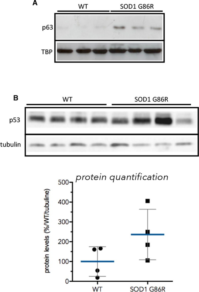
(A) Proteins from muscles were immuno-precipitated with a p63 antibody and then separated on a 10% SDS PAGE gel. Western blot experiment was performed using an antibody against p63 total. Shows pools of proteins from 3 animals at 105d. TBP was used as loading control. (B) Proteins (40 µg) from muscles were separated on 10% SDS PAGE gel. Western blot probing was performed with p53 antibody (IC12, 1/2000, Cell Signaling, Danvers, MA) and True Blot (Rockland Immunochemicals, Pottstown, PA) secondary antibody avoiding Ig heavy chain recognition. Tubilin was used as loading control. Graph below shows% of induction relative to the mean of p53 expression level in WT animals normalised with tubulin.
Figure 4—figure supplement 2. Gastrocnemius muscles from wild-type or symptomatic SOD1(G86R) (105 days) mice were cryodissected and probed for total p73 protein.

Graph represents the number of fibers per surface unit as indicated (n = 5). *p<0.01 compared to control, as calculated by a one-way ANOVA test followed by a Tukey post-test.
Figure 4—figure supplement 3. Gastrocnemius muscles from wild-type or symptomatic SOD1(G86R) (105 days) mice were cryodissected and probed for total p63 protein and nuclei (Hoechst).
