(
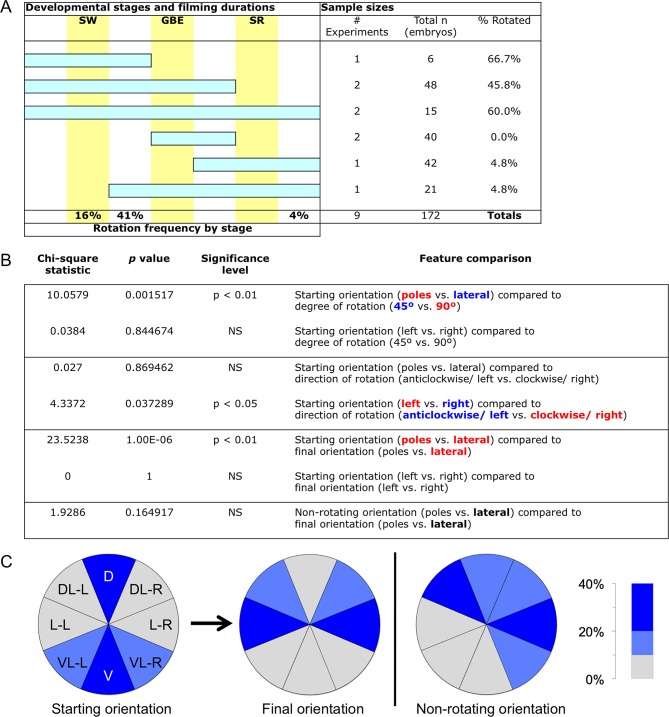
A) Sample sizes and film durations (blue bars) relative to landmark developmental stages (yellow bars: SW, serosal window closure; GBE, germband extension complete; SR, serosal rupture; staging as in
Koelzer et al. (2014)). Embryos were analyzed from multiple time-lapse data sets of at least 15.75-hr duration at 30°C, recorded with an inverted DeltaVision RT (Applied Precision) microscope. Pooled data included embryos in the nGFP and various enhancer trap backgrounds (
G04609-heart,
G12424-serosa, KT650-serosa, HC079-amnion), including heterozygote crosses of HC079 with each of
G04609,
G12424, and nGFP. (Background autofluorescence was sufficient for scoring early development in the absence of specific GFP signal.) 'Frequency by stage' refers to the beginning of rotation; all embryos beginning during (16%) or after (41%) the SW stage completed rotation before the GBE stage. Rarely, a moderate degree of rotation occurred during dorsal closure (4%). (
B). Chi-square statistics for specified comparisons. For significant results, text color-coding indicates the nature of the interaction. Longitudinal rotation ranged from approximately 20 to 100 degrees, with two-thirds of embryos rotating approximately 90°. Embryos laying on their dorsal or ventral surface ('poles') were more likely (69%) to rotate 90°, while embryos in lateral aspect (85%) tended to only rotate 45°. While overall there was no bias in direction of rotation, embryos initially laying on their right sides tended to rotate anticlockwise (70%) while embryos on their left sides rotated clockwise (78%). Direction of rotation was from the perspective of the embryo’s posterior (
i.e., clockwise is right, anticlockwise is left). These directional biases are applicable across the full range from ventral-lateral through dorsal-lateral, such that rotation tended to result in more strictly lateral final orientations (64%). Meanwhile, most non-rotating embryos began in a lateral orientation (89%). (
C) Pie charts representing the eight 45°-sectors of the egg circumference (D, dorsal; DL-L and -R, dorsal-lateral-left and -right; L-L and -R, lateral-left and -right; VL-L and -R, ventral-lateral-left and -right; V, ventral), color-coded for frequency of occurrence according to the heat map.


