Figure 4. Increasing p-eIF2α in young mice blocks cocaine-induced LTP and behavior.
(a) Schematic showing Sal003 mechanism of action. (b–c) Infusion of Sal003 into the VTA blocked cocaine-induced potentiation (c, p<0.001, n=5 per group, t8=3.81) and increased p-eIF2α in the VTA (p<0.01, n=7/6 vehicle/Sal003, t11=3.172). (d) Schematic of experimental design. (e) Direct application of cocaine (1 μM) induced LTP 3–5 hr post-treatment (p<0.05, n=11/6 vehicle/cocaine, F2,20=7.48), whereas Sal003 prevented it (p<0.05, n=11/6, vehicle/cocaine+Sal003, F2,20=7.48, cocaine vs. cocaine+Sal003). Representative traces of AMPAR and NMDAR EPSCs (top). (f) Infusion of Sal003 into the VTA blocked CPP (p<0.05, n=7 vehicle+cocaine, t12=2.592; p=0.1147, n=10 Sal003+cocaine, t18=1.892) in adolescent mice. (g) Application of Sal003 (20 μM, 10 min), a selective inhibitor of eIF2α phosphatases, induced LTD in VTA DA neurons from adolescent mice (p<0.001, n=6, t10=9.517). Plots are mean ± s.e.m.

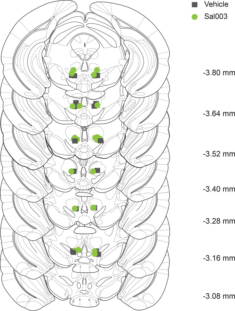
Figure 4—figure supplement 1. Sites of Sal003 infusions into VTA at seven rostrocaudal planes and corresponding increase in p-eIF2α. .