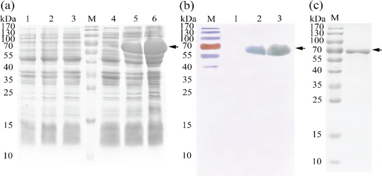
Fig. 6.
Expression of recombinant PmHsp60 in E. coli and western blot analysis. a. SDS-PAGE of expressed protein. Lanes M, pre-stained molecular weight marker; 1, non-induced cells transformed empty plasmid; 2, 3-h-induced cells transformed empty plasmid; 3, 6-h-induced cells transformed empty plasmid; 4, non-induced cells transfected recombinant; 5, 3-h-induced cells transformed recombinant; 6, 6-h-induced cells transformed recombinant. b. Western blotting of PmHsp60. Lanes 1, non-induced; 2, 3-h-induced PmHsp60-6 × His fusion protein; 3, 6-h-induced PmHsp60-6 × His fusion protein. c. purified PmHsp60-6 × His fusion protein

