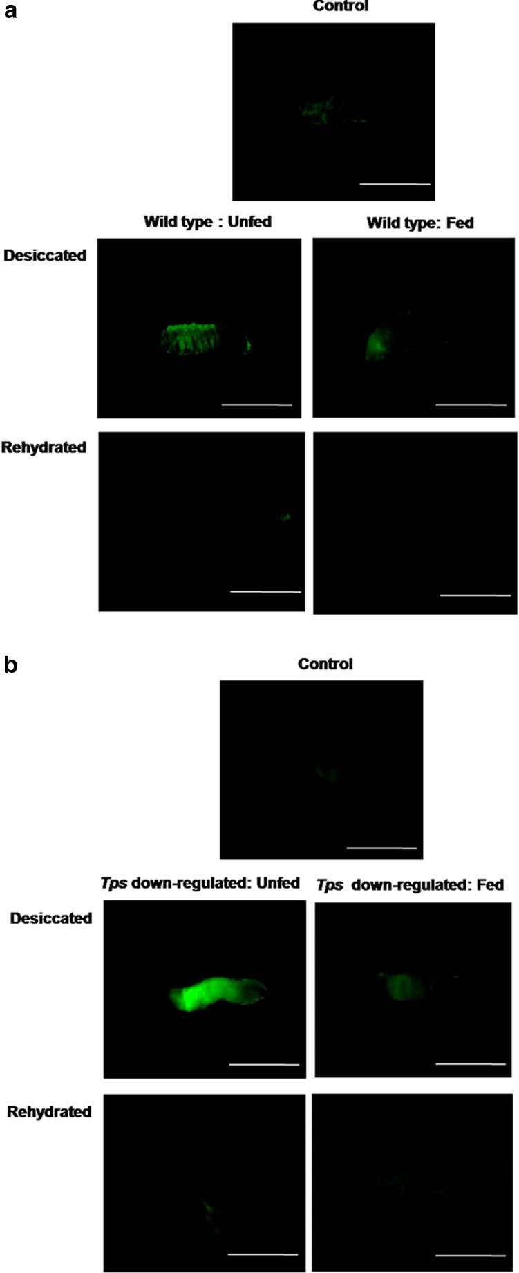
Fig. 4.
Representative images of whole larval live imaging under a fluorescence microscope. a Visualisation of O2 ·− using DCF-DA dye in the control, unfed and trehalose-fed wild-type larvae. b Visualisation of O2 ·− using DCF-DA dye in the control, unfed and trehalose-fed dTps1-downregulated larvae. Bar = 1 mm

