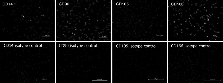
Fig. 4.
Immunofluorescence in CD90+ isolated cells from MPB after thawing at 15 days of monolayer culture, for superficial cell markers CD14, CD90, CD105, and CD166. Cell nuclei present as blue when stained with 4′ 6-diamidino-2-phenylindole [DAPI], and green with fluorescein isothiocyanate (FITC) fluorochrome-labeled antibodies. (Color figure online)

