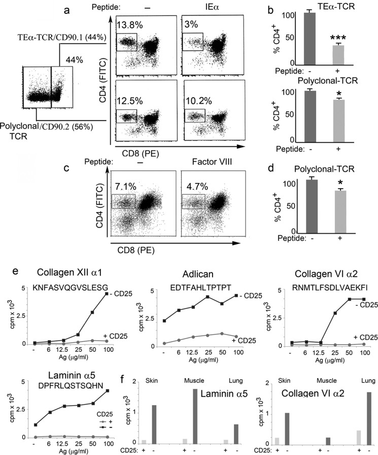
FIGURE 10.
Role of lymph peptides in central and peripheral tolerance. a, FACS analysis of thymocytes from chimeric mice generated by irradiating wild type C57Bl6 mice and reconstituting them with a mixture of bone marrow from TEα TCR transgenic mice (CD90.1) and C57Bl6 mice (CD90.2) in a 1:1 ratio. The percentage of CD4+ thymocytes is reported before and after injection of the I-Ab-restricted ASFEAQGALANIAVDK peptide. CD4+-CD90.1+ thymocytes derive from wild type C55Bl6 mice, whereas CD4+-CD90.2+ thymocytes derive from TEα TCR transgenic mice. b, bar graph showing the number of CD4+-CD90.1+ and CD4+-CD90.2+ thymocytes as determined in a. Mean ± S.D. of five separate experiments; ***, p < 0.01 for the depletion of TEα-TCR; *, p < 0.05 for the depletion of polyclonal TCR. c, FACS analysis of thymocytes from factor VIII knock-out mice. The number of CD4+ thymocytes is reported before and after injection of the I-Ab-restricted SPSQARLHLQGRTNAWRPQVNDPKQWLQVD peptide. d, bar graph of the percentage of CD4+ thymocytes, as determined in a. Mean ± S.D. of five separate experiments, *, p < 0.05 for the depletion of polyclonal TCR. e, nodal T cell proliferation of CD25+-competent or -depleted HLA-DR1+ mice. Inguinal lymph node were harvested and cultured for 4 days with titrated amounts of the reported peptides. f, T cell proliferation of CD25-competent or CD25-depleted HLA-DR1+ mice. Skin, muscle, and lung tissues were harvested and cultured for 4 days with titrated amounts of the same peptides.

