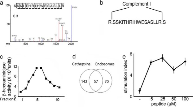
FIGURE 6.
Analysis of complement 3 processing. a, sequence and MS/MS spectrum of the complement 3 peptide found in the human lymph. b, cleavage sites by complement 1 processing. c, β-hexosaminidase activity in fractions collected from a 27/10 Percoll gradient to identify endosomal compartments. d, Venn diagram of the total number of complement 3 peptides sequenced following processing by recombinant cathepsins or endosomal compartments. e, T cell proliferation in response to the complement 3 peptide following immunization of HLA-DR1 transgenic, I-A, I-E, and C3 knock-out mice.

