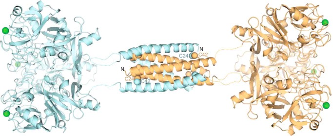
FIGURE 10.
Predicted structure of full-length XEEL. The ligand-binding site on each carbohydrate-binding domain (derived from the crystal structure) is marked by its calcium ion (green). The predicted structure of the dimerization domain (from PDB ID: 2SIV (62)) is shown as a helical bundle, with two of the six Cys-24–Cys-42 disulfide bonds labeled.

