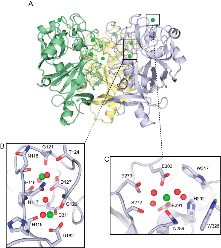
FIGURE 2.
XEEL is a trimer with multiple calcium ion binding sites. A, crystal structure of Se-Met-labeled XEELCRD trimer oriented with the intelectin-specific domains toward the top of the figure and the fibrinogen-like lobes below. The second trimer in the asymmetric unit is removed for clarity. B, structural calcium site with two calcium ions (green) and four ordered water molecules (red). C, ligand-binding site with one calcium ion (green) and four ordered water molecules (red).

