FIGURE 1.

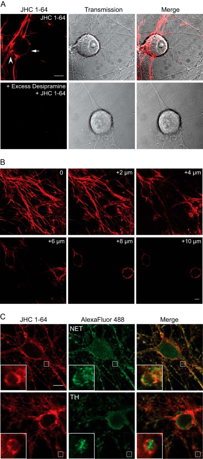
Visualization of endogenously expressed NET in live cultured sympathetic neurons using the fluorescent cocaine analogue JHC 1-64. A, labeling of JHC 1-64 is blocked by the NET inhibitor desipramine (100 μm). The SCG neurons were incubated with 50 nm JHC 1-64 (top) or preincubated with 100 μm desipramine (bottom) for 20 min before incubation for 20 min with 50 nm JHC 1-64. The experiments shown are representative of at least three similar experiments with the same results. Scale bar, 10 μm. B, z-scan of JHC 1-64-labeled SCG neurons. The scan demonstrates uniform surface distribution of NET in the axonal network, boutons, and somata. The pictures represent single confocal sections starting from the bottom of the neurite tree (0) and ending on 10 μm above the surface, on the level of the neuronal somas. The neuronal cultures were incubated with 50 nm JHC 1-64 before the analysis was performed. C, JHC 1-64 labels SCG neurons immunopositive for NET and tyrosine hydroxylase (TH). Cultured SCG neurons were labeled with 50 nm JHC 1-64 for 20 min before neurons were fixed, permeabilized, and stained with antibodies against the noradrenergic cell marker NET (top) or tyrosine hydroxylase (bottom). Costaining of SCG neurons with JHC 1-64 and mouse anti-NET antibody showed extensive overlap in the extensions and in the plasma membrane of the soma as well as the boutons. Costaining of SCG neurons with JHC 1-64 and anti-tyrosine hydroxylase antibody showed labeling of same cells but no direct overlap because the majority of tyrosine hydroxylase staining appeared intracellular. The experiments shown are representative of at least three similar experiments with the same results. The small white squares mark areas that are shown enlarged inside large squares. Scale bar, 10 μm.
