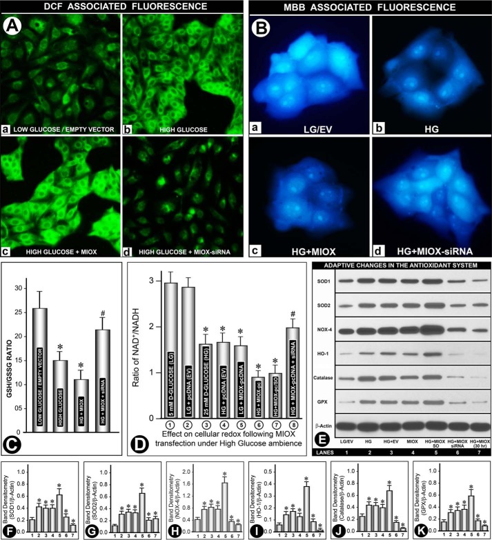
FIGURE 4.
A, effect of HG on ROS generation following MIOX transfection. A low level DCF-associated green fluorescence was observed under LG in the presence or absence of empty vector (A (a)). HG induced a robust increase in fluorescence, which was further accentuated with transfection of MIOX-pcDNA (A (b and c)). Fluorescence was markedly reduced with MIOX siRNA treatment (A (d)). B, effect of HG on the relative depletion of cellular GSH following MIOX transfection. A bright MBB-associated blue fluorescence was observed under LG ambience in the presence or absence of EV (B (a)). HG significantly reduced fluorescence, which was further decreased with the transfection of MIOX-pcDNA (B (b and c)), and it was restored with treatment with MIOX siRNA (B (d)). In addition, the GSH/GSSG ratio was also determined (C). Under HG, the ratio was decreased, and a further decrease was seen in cells overexpressing MIOX. MIOX siRNA treatment prevented such a decrease to a large extent. D, perturbation in NAD+/NADH ratio following MIOX transfection in HG ambience. About a 50% decrease in the NAD+/NADH ratio was observed under HG ambience compared with LG concentration, and no further decrease was seen with transfection of EV (columns 1–4). A comparable decrease in the ratio was observed following MIOX-pcDNA transfection alone under LG ambience, and it further decreased in the presence of HG ambience (columns 5 and 6). Treatment with siRNA improved the NAD+/NADH ratio in MIOX-transfected cells maintained under HG ambience, whereas scramble oligonucleotide had no significant effect (columns 7 and 8). E, profile of antioxidant defense system and oxidant-responsive enzymes in response to perturbation in the intracellular redox. Western blotting analyses of antioxidant enzymes (i.e. SOD1, SOD2, NOX4, heme oxygenase-1 (HO-1), catalase, and glutathione peroxidase (GPX)) revealed a variable increase in their expression up to 15 h of the culture period under HG ambience. Concomitant transfection with EV did not cause any further increase in their expression, as compared with that observed at LG concentration (lanes 1–3). Transfection with MIOX-pcDNA yielded a comparable increase, and it was notably increased under HG ambience, whereas it was reduced to basal levels by MIOX siRNA treatment (lanes 4–6). Interestingly, the expression of various enzymes of the scavenging system was reduced in the 30 h time frame of the culture. Band densities normalized to respective β-actin band densities of various blots of each variable (i.e. SOD1, SOD2, NOX4, heme oxygenase-1, catalase, and glutathione peroxidase) compared with their respective controls at the 15 h time point are shown in bar graphs in corresponding panels F–K (n = 4; *, p < 0.01; #, p < 0.05). Error bars, S.D.

