FIGURE 6.
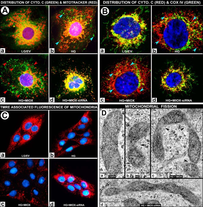
A and B, effect of MIOX overexpression on mitochondrial dysfunction with release of cytochrome c under HG ambience. Under LG ambience, the cytochrome c (green) and Mitotracker (red) co-localized to mitochondria and yielded a deep orange/yellow-colored fluorescence (A (a)). Treatment with HG led to a moderate leakage of cytochrome c into the cytosolic compartment (A (b), bluish arrowheads). The MIOX-pcDNA transfection under HG ambience led to leakage of cytochrome c into the cytosol (A (c), red arrowheads). The siRNA treatment of MIOX-pcDNA-transfected cells maintained under HG ambience restored the co-localization of cytochrome c and Mitotracker, yielding a yellow-colored fluorescence (A (d)). Cytochrome c release experiments were also performed using another mitochondrial marker, COX IV, which does not dissociate from mitochondria under oxidant stress. Under low glucose conditions, cytochrome c (red) and COX IV (green) co-localized in the mitochondria and yielded a yellow-colored fluorescence (B (a)), and HG treatment led to a moderate degree of leakage of cytochrome c (B (b), bluish arrowheads), whereas MIOX transfection under HG ambience led to a notable dissociation of cytochrome c into the cytosol (B (c), bluish arrowheads), and siRNA treatment largely restored the co-localization of cytochrome c and COX IV, yielding a yellow-colored fluorescence (B (d)). C, effect of MIOX overexpression on ΔΨm under HG ambience. Under LG ambience, TMRE staining is seen uniformly localized to the mitochondria (C (a)). A moderate loss of TMRE staining was observed with the treatment with HG (C (b)). Transfection of cells with MIOX-pcDNA under HG ambience caused a loss of staining (i.e. loss of ΔΨm) (C (c)), whereas siRNA treatment restored the cellular TMRE fluorescence to basal levels (C (d)). D, effect of MIOX overexpression on mitochondrial ultrastructure under HG ambience. By electron microscopy, cells maintained in medium containing LG showed normal morphology in the majority (∼70%) of mitochondria (>1 μm in length along the long axis) with intact inner and outer membranes and cristae (D (a)). Under HG ambience, the population of tubular mitochondria decreased to 47%, and there were constrictions in the body of the mitochondria accompanied by a relative loss of their cristae (D (b), arrowhead). Overexpression of MIOX further decreased the population of intact mitochondria to 35% and induced marked constrictions and at times U-shaped angulation or other deformations of mitochondria in the presence of HG ambience (D (c), arrowheads), suggesting that mitochondria are undergoing a significant degree of fission. MIOX siRNA treatment significantly lessened the constrictions, and a majority (∼63%) of mitochondria regained their tubular morphology (D (d)).

