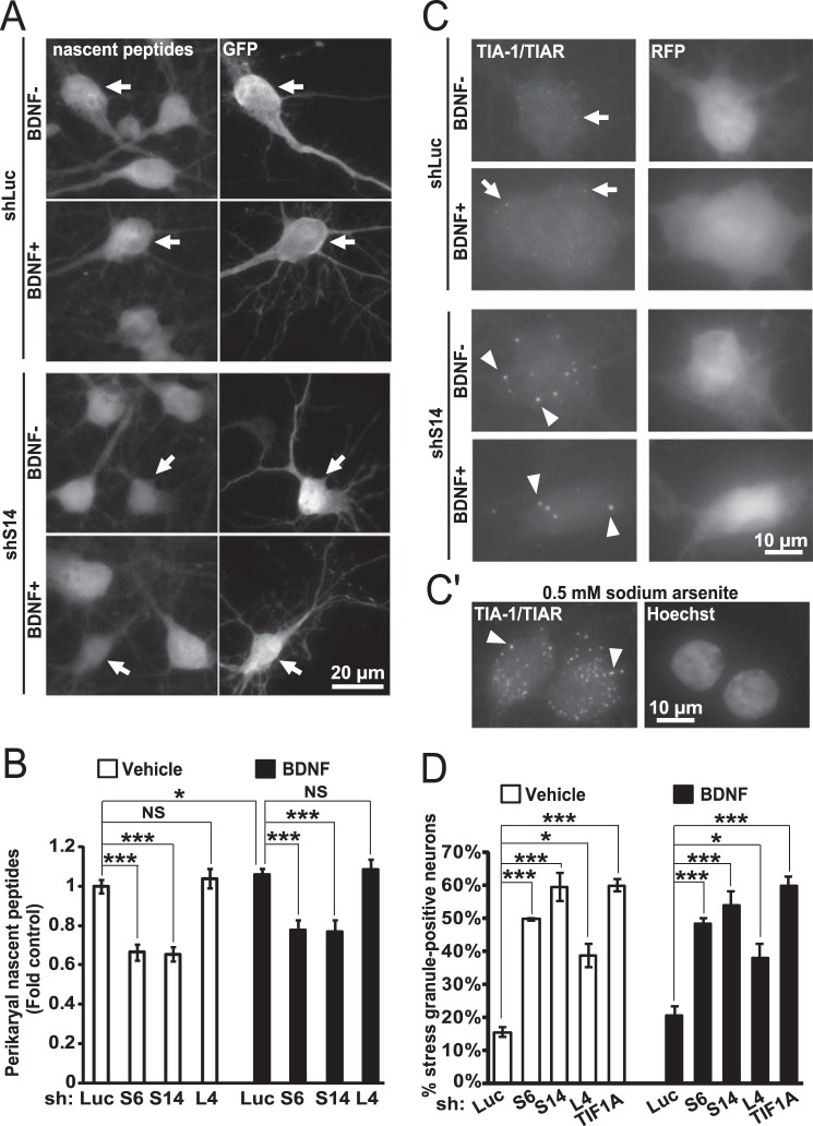
FIGURE 7.
Impairment of global protein synthesis after inhibition of ribosomal biogenesis. A and B, DIV6 hippocampal neurons were transfected as for Fig. 5C. After 3 days, cells were treated with 50 ng/ml BDNF. After 45 min, OPP was added followed by cell fixation 30 min later. The OPP-labeled nascent peptides were visualized with fluorescent Click-iT chemistry. A, representative images depicting nascent peptide accumulation in shLuc- and shS14-transfected neurons (arrows). B, nascent peptide accumulation in the perikarya was reduced by shS6 or shS14 but not shL4 (two-way ANOVA, effect of shRNA, F3,459 = 49.425, p < 0.01). Data represent means ± S.E. of at least 50 individual cells from three independent experiments; *, p < 0.05; ***, p < 0.001 (post hoc tests). C and D, DIV6 hippocampal neurons were transfected as for Fig. 5, D and E. After 2 days, 50 ng/ml BDNF was added for 24 h as indicated. SGs were visualized by TIA-1/TIAR immunostaining. C, representative images of shLuc and shS14-transfected neurons. In shLuc-transfected neurons TIA-1/TIAR signal is visible throughout the soma, including fine extra-nuclear granules (arrows). In shS14-transfected cells, the diffuse signal is reduced, and large granules appear in the perikaryal region and proximal dendrites (arrowheads) consistent with SG morphology. C′, well established inducer of SGs, sodium arsenite (0.5 mm), was added to neuronal cultures for 1 h. Note similarity of arsenite-induced SGs to those produced after depletion of S14 (arrowheads). D, fraction of SG-positive cells increased when ribosomal biogenesis was inhibited by knockdowns of RPs or TIF1A. Such a response was also present in BDNF-treated neurons. NS, not significant. Data represent means ± S.E. of three independent experiments; ***, p < 0.001, U test.

