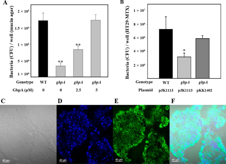
FIGURE 1.
Effect of GbpA on mucin binding and host cell adhesion of V. vulnificus. Upper panel, mucin binding activities of the strains. A, strains (∼107 cfu) were added to each well of 12-well culture dishes containing the mucin-agar and various amounts of GbpA provided exogenously as indicated. After 1 h of incubation, the adherent bacterial cells were enumerated as cfu per well. WT, wild type; gbpA, gbpA mutant. B, mucin-secreting HT29-MTX cells (∼107 cells) seeded in each well of 12-well culture dishes were infected at a multiplicity of infection of 10 with the strains as indicated. After a 30-min incubation, the adherent bacterial cells were enumerated as cfu per well. Error bars represent the S.D. *, p < 0.05, and **, p < 0.005 relative to the wild type. WT (pJK1113, empty vector), wild type; gbpA (pJK1113), gbpA mutant; gbpA (pKK1402), complemented strain. Lower panel, development of the mucin-secreting HT29-MTX cells. C, bright field image of HT29-MTX cells. D, nucleus of HT29-MTX cells was stained blue with DAPI. E, mucin of HT29-MTX cells was stained green with the anti-MUC5AC primary antibody and then labeled with FITC-conjugated secondary antibody. F, merged image of C–E. Images are visualized using a confocal laser scanning microscope. Scale bar, 40 μm.

