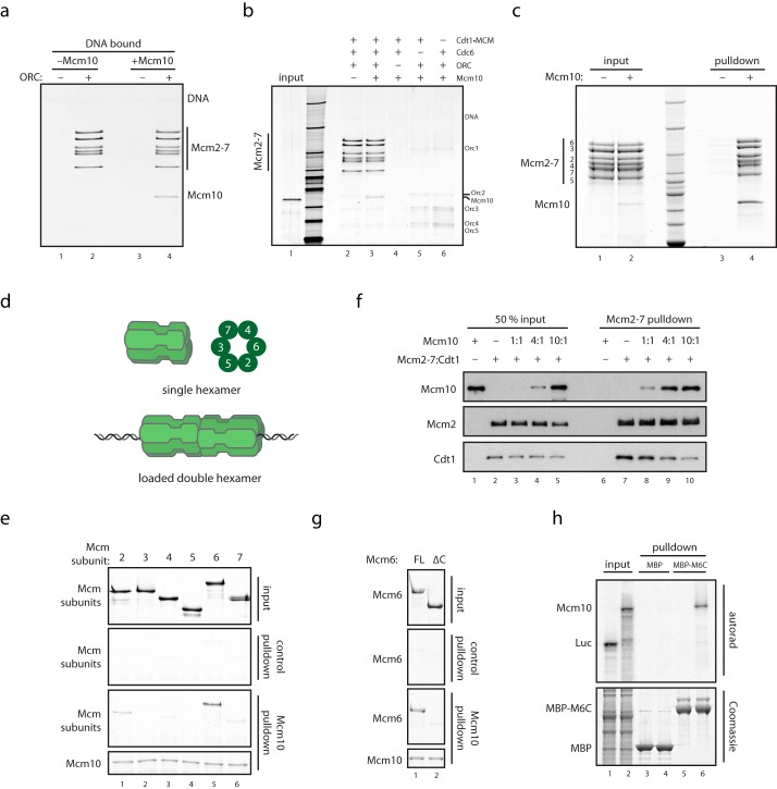
FIGURE 1.
Mcm10 binds directly to MCM via Mcm2 and Mcm6. a, purified MCM was loaded onto DNA with purified loading factors and washed with high salt to remove ORC and Cdc6, and DNA-bound double hexamers were mixed with Mcm10 and analyzed for Mcm10 recruitment by SDS-PAGE and silver stain. b, MCM loading reactions were carried out as described (14), with individual loading factors excluded as indicated. Reactions were washed with low salt buffer, mixed with Mcm10, and washed, and DNA-bound proteins were analyzed by SDS-PAGE and silver stain. c, purified Mcm10 containing an N-terminal His tag was mixed with purified single hexamers of MCM. The sample was bound to Ni-NTA resin, which was washed and analyzed by SDS-PAGE and InstantBlue stain. d, schematic showing soluble MCM single hexamers, MCM double hexamers loaded onto DNA, and the order of subunits within the MCM ring. e, subunits of MCM were purified after overexpression in bacteria and mixed with anti-T7 resin bound to N-terminally T7-tagged Mcm10. Resin was washed, and bound proteins were analyzed by SDS-PAGE and InstantBlue stain. f, purified Mcm10 was mixed with a stoichiometric complex of MCM·Cdt1; MCM was immunoprecipitated with anti-FLAG resin via a 3×FLAG tag on Mcm3. Resin was washed, and bound proteins were analyzed by SDS-PAGE and Western blotting. g, full-length (FL) Mcm6 and Mcm6(1–871) purified after overexpression in bacteria were mixed with anti-T7 resin bound to N-terminally T7-tagged Mcm10. Resin was washed, and bound proteins were analyzed by SDS-PAGE and InstantBlue stain. h, MBP and MBP-Mcm6(789–1017) bound to glutathione-Sepharose resin were mixed with rabbit reticulocyte lysate containing 35S-labeled Mcm10 or luciferase (Luc). Resin was washed, and bound proteins were separated by SDS-PAGE and analyzed with InstantBlue stain or autoradiography.

