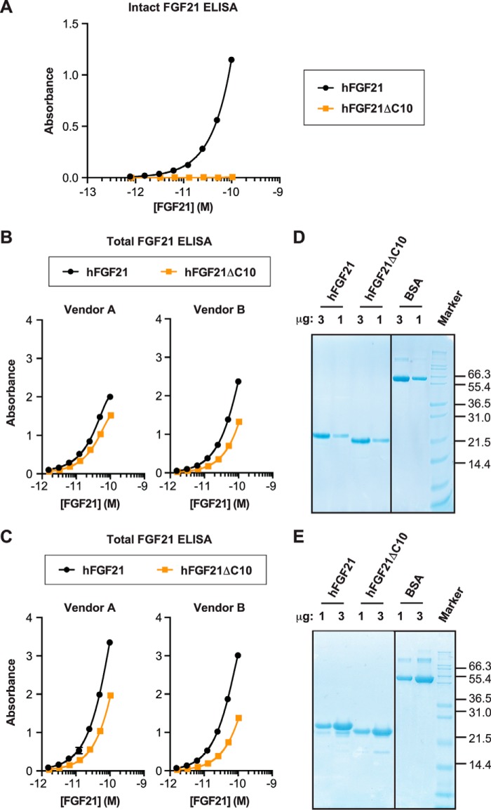
FIGURE 4.
Evaluation of FGF21 ELISA. A and B, wild-type hFGF21 and the FAP-digested derivative (hFGF21ΔC10) were tested by using intact hFGF21 ELISA (A) or total hFGF21 ELISA from two separate sources (B). C, hFGF21 (L98R) and the FAP-digested derivative (hFGF21ΔC10) were tested by using the total hFGF21 ELISA used in B. A–C, each protein was tested in duplicate at each concentration, and the results are plotted as mean ± S.E. D and E, Coomassie staining of SDS-PAGE gels. The proteins used for the ELISAs in B and C were analyzed in D and E, respectively, with BSA as a control.

