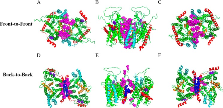
FIGURE 1.
Placement of pBpA at the dimer interface for in vivo photo-cross-linking. A–C, two protomers of Thermotoga maritima SecYEG were manually docked into a front-to-front conformation based on cysteine cross-linking studies (16, 17). SecY, SecE, and SecG are colored green, red, and cyan, respectively. TM2b and 7 of the lateral gate are colored orange. Sites with amber mutations for incorporation of pBpA are shown as magenta spheres. Views from the A, cytoplasm; B, along the plane of membrane; and C, periplasm are shown. D–F, similar to A–C except protomers were docked into a back-to-back conformation. Magenta and blue spheres indicate the amber mutations on SecY and SecE subunits, respectively. Two negative controls, shown in purple spheres, are amber mutations engineered into regions distant to either dimer interface.

