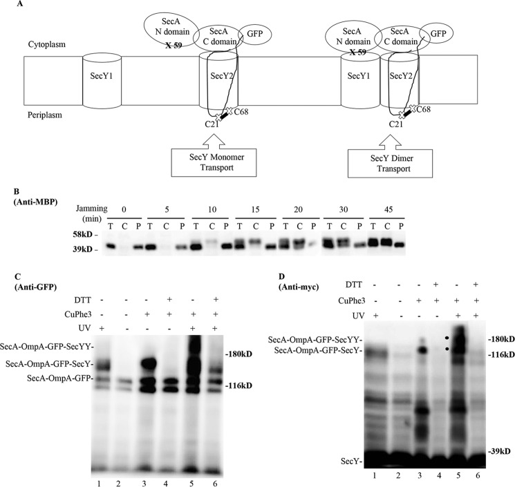
FIGURE 11.
SecA footprinting study of SecY dimer status. A, schematic of SecA footprinting study, where interaction of the NBD-1-NBD-2 (N domain) half of SecA with SecY1 or PPXD-HSD-THF-HWD-CTL (C domain) half of SecA with SecY2 are depicted along with the OmpA-GFP chimera attached to SecA. Sites for photo-cross-linking the SecA N domain (59) with SecY1 or for disulfide cross-linking the OmpA signal sequence (C21) to the plug domain (C68) of SecY2 are shown. B-D, the secY Cys68 mutant carrying the pBAD-secA-OmpA-GFP plasmid with the ompA Cys21 and secA 653 Amber (panel B) or secA 59 Amber (panels C and D) allele was grown in the presence of 1 mm pBpA and 30 μm IPTG until A600 reached 0.15, when arabinose was added to a final concentration of 0.2%. 45 min later, cells were harvested and either fractionated (panel B) or subjected to cross-linking (panels C and D). B, Western blot of cells divided into cytoplasm/membrane (C) and periplasmic (P) fractions, compared with total (T) cell input, probed with MBP antibody. C and D, Western blot of isolated membranes from cells treated with UV irradiation, CuPhe3 and/or DTT as indicated probed with GFP or c-Myc antibody as indicated. A large degradation fragment is visible immediately below the SecA-OmpA-GFP trimera.

