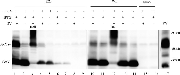
FIGURE 2.
Verification of in vivo photo-cross-linking. Each strain was grown as indicated in the presence or absence of 1 mm pBpA until A600 reached 0.3, when SecYEG was induced or not with IPTG at a final concentration of 1 mm for 2 h. Cells were harvested, resuspended in PBS buffer, and treated with UV irradiation (350–365 nm) for 20 min where indicated. Cell membranes were isolated and analyzed by Western blotting using c-Myc antibody as described under “Experimental Procedures.” Samples in lanes 3 and 12 were heated at 100 °C for 5 min prior to loading on the gel to induce SecY aggregation. YY indicates a genetically fused SecY dimer used as a marker (48). Anti-SecY peptide antiserum was used to detect this latter species.

