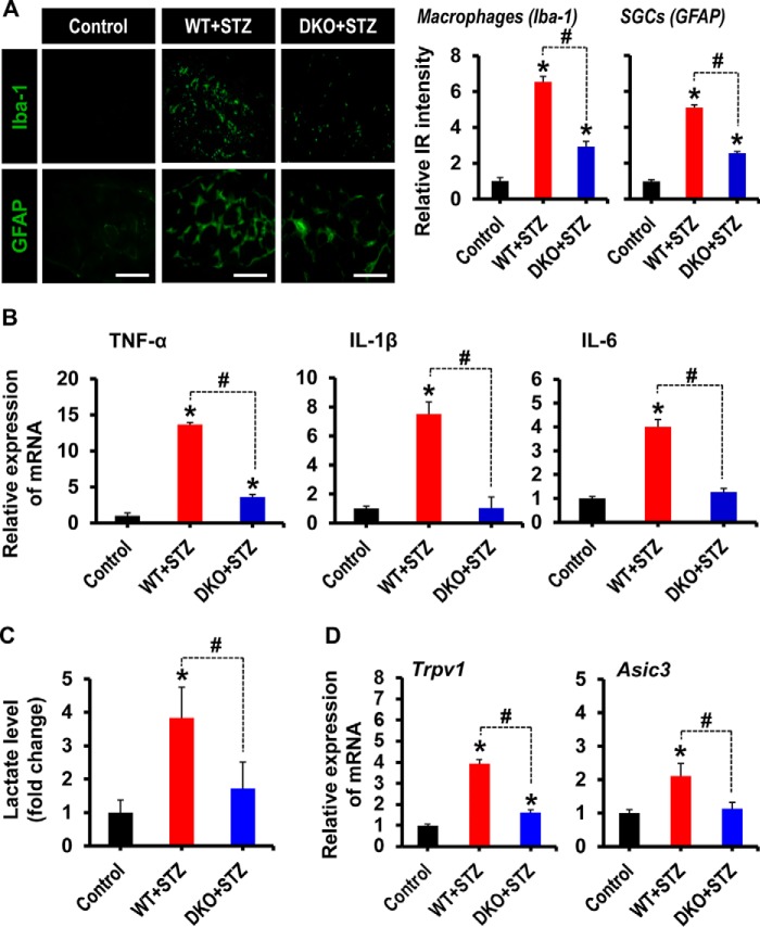
FIGURE 4.
Pdk2/4 deficiency attenuates macrophage infiltration, activation of satellite glial cells, proinflammatory cytokines expression, lactate surge, and expression of Trpv1 and Asic3 in the diabetic DRG. A, Iba-1(a macrophage marker) and GFAP (an SGC marker) immunostainings reveal their increased immunoreactivity in the DRG of WT animals at 3 weeks post-STZ injection, whereas Pdk2/4 deficiency significantly attenuates such an increase in immunoreactivities. Quantifications and statistical analyses of stained images are presented in adjacent graphs. Scale bars, 100 μm. *, p < 0.05 versus the vehicle-treated control animals; #, p < 0.05 between the indicated groups, Student's t test, n = 6; mean ± S.E. IR, immunoreactivity. B, the relative expression of TNF-α, IL-1β, and IL-6 mRNAs in the DRGs after 3 weeks of STZ injection as evaluated by real-time RT-PCR. C, lactate assay was performed to measure the lactate accumulation in the DRG at 3 weeks after STZ injection. Results of lactate levels are presented as the -fold change relative to control. D, the expression of Trpv1 and Asic3 mRNAs in the DRGs at 3 weeks after STZ injection was assessed by real-time RT-PCR. Results for mRNA expression are displayed as the -fold increase of gene expression normalized to GAPDH. *, p < 0.05 versus the vehicle-treated control animals; #, p < 0.05 between the indicated groups, Student's t test, n = 6 (for B and D) or n = 3 (for C); mean ± S.E. (error bars).

