
FIGURE 8.
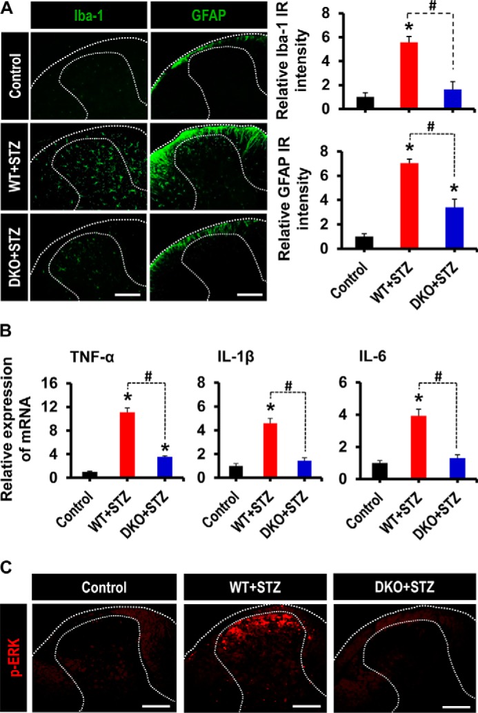
Role of PDK2/4 in glial activation, expression of proinflammatory cytokines, and phosphorylation of ERK in the spinal cord of diabetic mice. A, Iba-1 (a microglia marker) and GFAP (an astrocyte marker) immunoreactivities significantly increased in the dorsal horn of the lumbar segment of the spinal cord of WT mice at 3 weeks post-STZ injection, whereas these immunoreactivities significantly diminished in the diabetic DKO mice. Dotted lines demarcate the white and gray matters in the spinal cord dorsal horn. Quantifications and statistical analyses of stained images are presented in adjacent graphs. B, the relative mRNA expression of TNF-α, IL-1β, and IL-6 in the lumbar segment of the spinal cord (L4–6) at 3 weeks after STZ injection as assessed by real-time RT-PCR. Results for mRNA expression are displayed as the -fold increase of gene expression normalized to GAPDH. C, immunofluorescence staining was performed to detect the expression of p-ERK in the lumbar segment of the spinal cord from vehicle/STZ-injected animals at 3 weeks post-injection. Scale bar, 200 μm. Images show the representative results of at least three independent experiments. *, p < 0.05 versus the vehicle-treated control animals; #, p < 0.05 between the indicated groups, Student's t test, n = 6 (for A and B) and n = 3 (for C); mean ± S.E. (error bars).