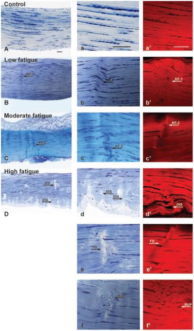
Figure 4.
Morphology of nonloaded, control, and fatigue damaged tendons under bright-field (A–D in low magnification, a–f in high magnification; stained in Toluidine blue) and confocal (a′ –f′ bulk stained in basic Fuchsin) microscopy. Both bright-field (A, a) and confocal (a′) microscopy exhibited parallel fibers that are highly aligned in nonloaded, control tendons. Tendons at the Low fatigue damage level are characterized by kinked fiber deformation (KF-1 in B, b, b′). At the Moderate fatigue damage level, tendons showed the same type of kinked fiber deformation patterns with an increase in the amount of damage area fraction (KF-2 in C, c, c′). Microstructural changes at the High fatigue damage level are characterized by: i) dissociation among fibers (DIS in D, d, d′); ii) transversely-oriented fiber discontinuities (FD in D, e, e′); and iii) fiber rupture patterns (RUP in f, f′; not shown in D). Scale bar=100 μm.

