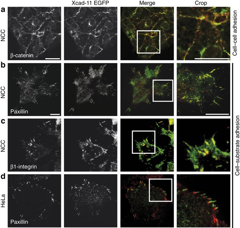
Figure 1. Xcad-11 is localized in focal adhesions.
Xenopus NCC injected with Xcad-11-EGFP, explanted on fibronectin-coated glass dishes and immunostained for (a) β-catenin, (b) paxillin and (c) β1-integrin. (a) A confocal image focused on the apical side of NCC shows co-localization of Xcad-11 with β-catenin at cell–cell contacts. (b,c) TIRF images demonstrating co-localization of Xcad-11 with paxillin and β1-integrin in focal adhesions at the cell substrate. (d) HeLa cells transfected with Xcad-11-EGFP, immunostained for paxillin and imaged by TIRF microscopy display partial localization of Xcad-11 with paxillin at the cell substrate. Scale bars, 20 μm (a); 10 μm (b–d).

