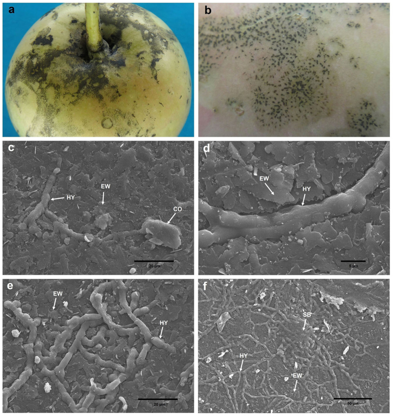
Figure 5. Photographs and scanning electron micrographs of P. fructicola on apple fruit.
(a,b) SBFS signs caused by P. fructicola on apple fruit. (c) Germinating conidium and primary hypha partly submerged into the surface of epicuticular waxes. (d,e) Hyphae partly submerged into the surface of epicuticular waxes. (f) Sclerotium-like body and hyphae, both of which are partly submerged into the surface of epicuticular waxes. CO, conidium; HY, hypha; EW, epicuticular wax; SB, sclerotium-like body.

