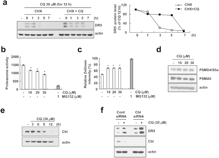
Figure 5. CQ induces DR5 up-regulation through sustained protein stability in Caki cells.
(a) Caki cells were treated 30 μM CQ for 12 h, then changed with fresh medium and pre-treated 20 μg/ml cycloheximide (CHX) for 30 min, and treated with or without 30 μM CQ for the indicated time periods. The protein expression of DR5 was determined by Western blotting (left panel). The band intensity of DR5 protein was measured using the public domain JAVA image-processing program ImageJ (http://rsb.info.nih.gov/ij) (right panel). (b) Caki cells were treated with 1 μM MG132 (as a positive control) and indicated concentrations CQ for 24 h. The cells were lysed, and the proteasome activity was measured as described in the Materials and Methods. (c) Caki/ZsGreen cells were treated with 1 μM MG132 (as a positive control) and indicated concentrations of CQ for 24 h. Proteasome activity was analyzed by using FACS analysis. (d) Caki cells were treated with indicated concentrations of CQ for 24 h. The protein levels of PSMD4/S5a and PSMA5 were determined by Western blotting. (e) Caki cells were treated with 30 μM CQ for the indicated time periods. The protein levels of Cbl was determined by Western blotting. (f) Caki cells were transfected with Cbl siRNA or control siRNA. Twenty-four hours after transfection, cells were treated with 30 μM CQ for an additional 24 h. The protein expression of DR5 and Cbl were determined by Western blotting. The level of actin was used as a loading control. The values in panel (b,c) represent the mean ± SD from three independent samples. *p < 0.05 compared to the control.

